Confocal.nl in the news
From Chile to the Netherlands: how Desiree started at Confocal.nl
Read more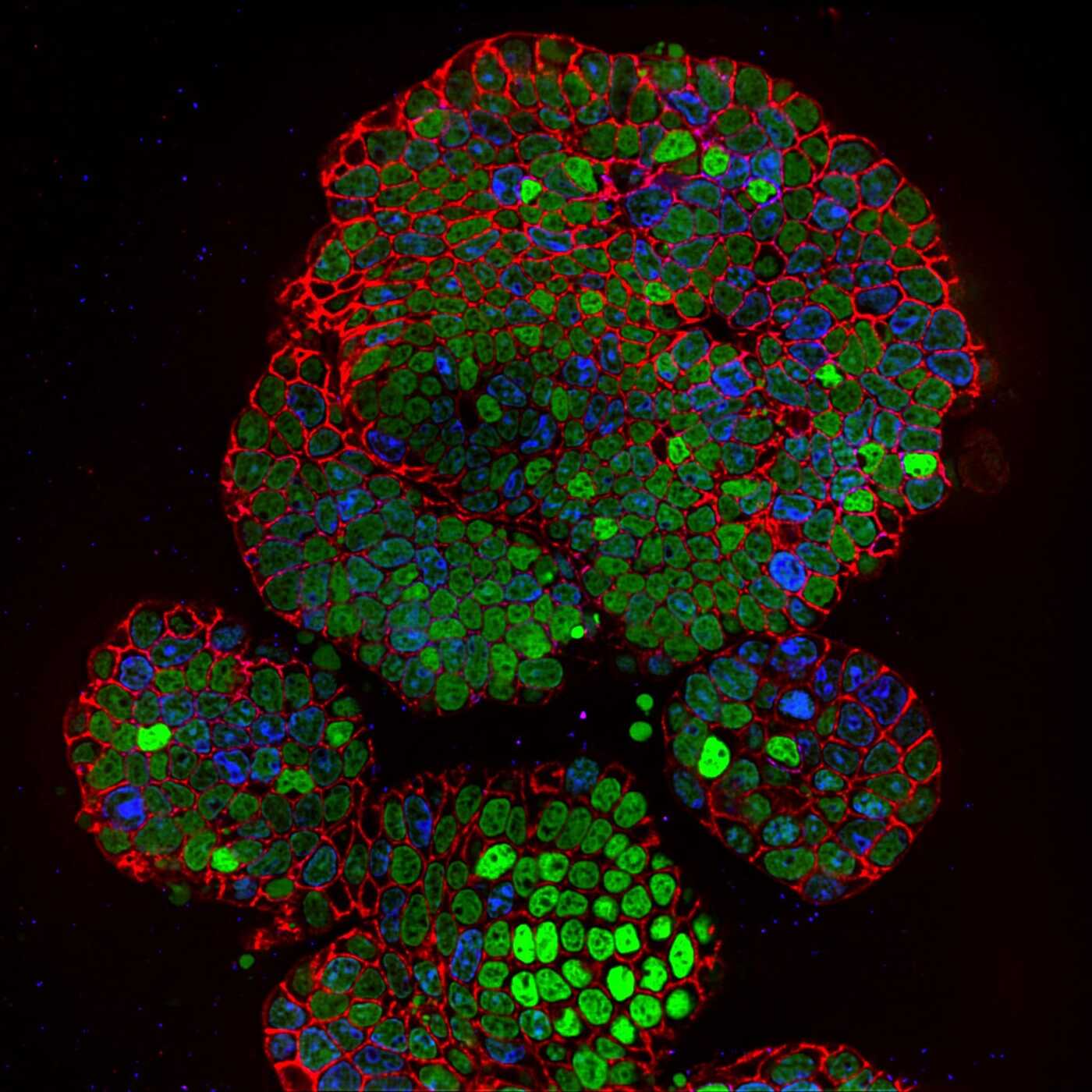
News
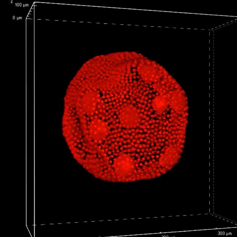
Line REscan confocal microscope NL5+: a fast and sensitive technique for studying organoids
Read more
Articles
How super-resolved fluorescence microscopy led to a Nobel Prize
Read more
News
Confocal.nl enters new growth phase with investor and partner ECFG
Read more


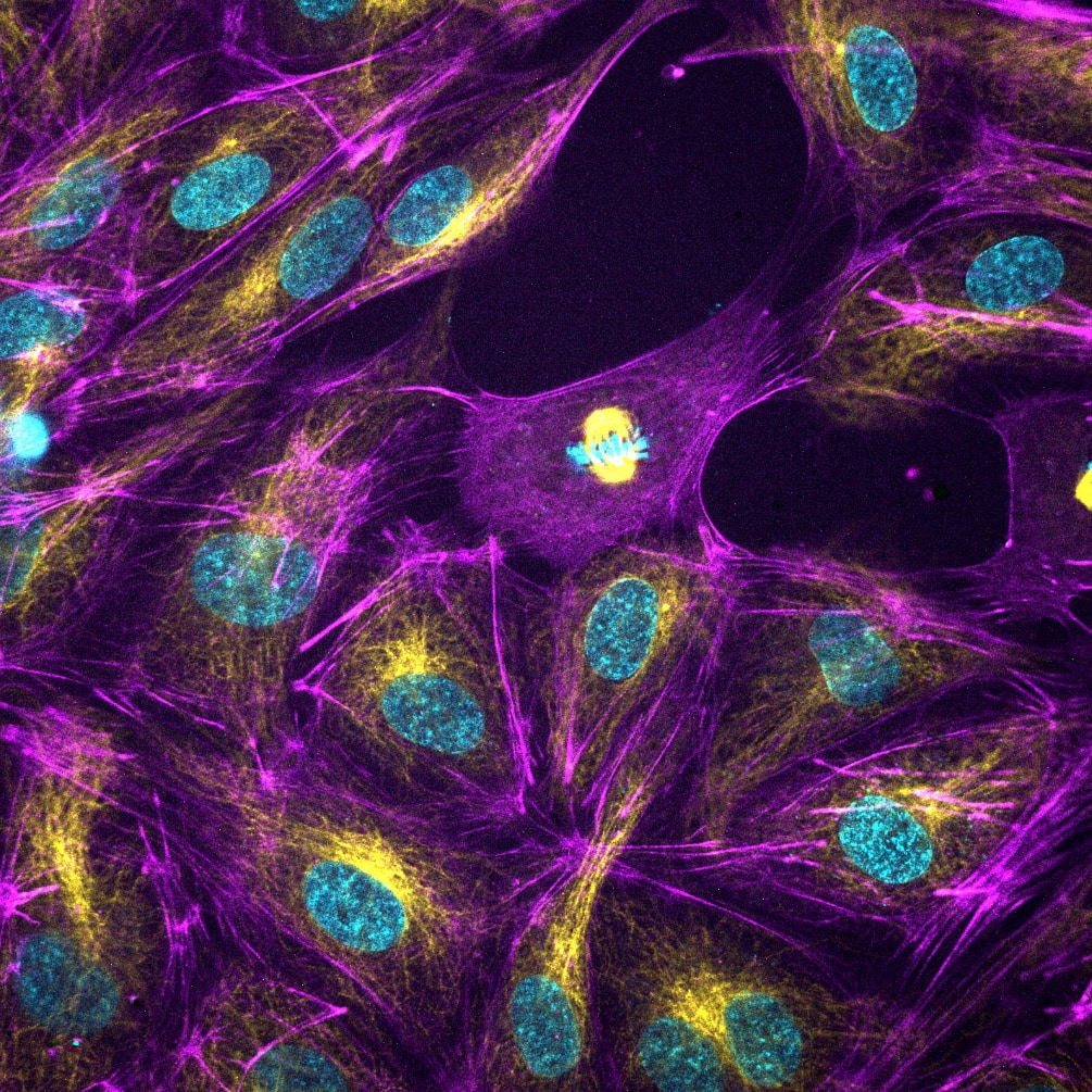
"RCM is a very easy to use system
that we have in combination with
STORM and it provides great
underlay images for STORM data."

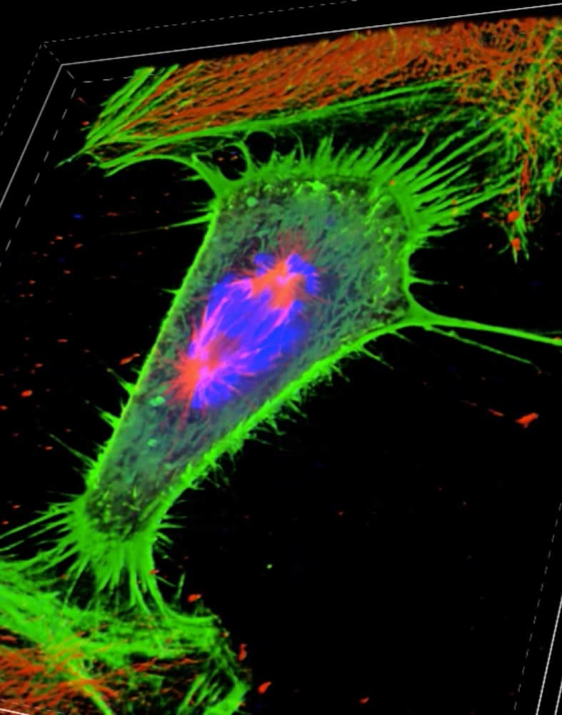
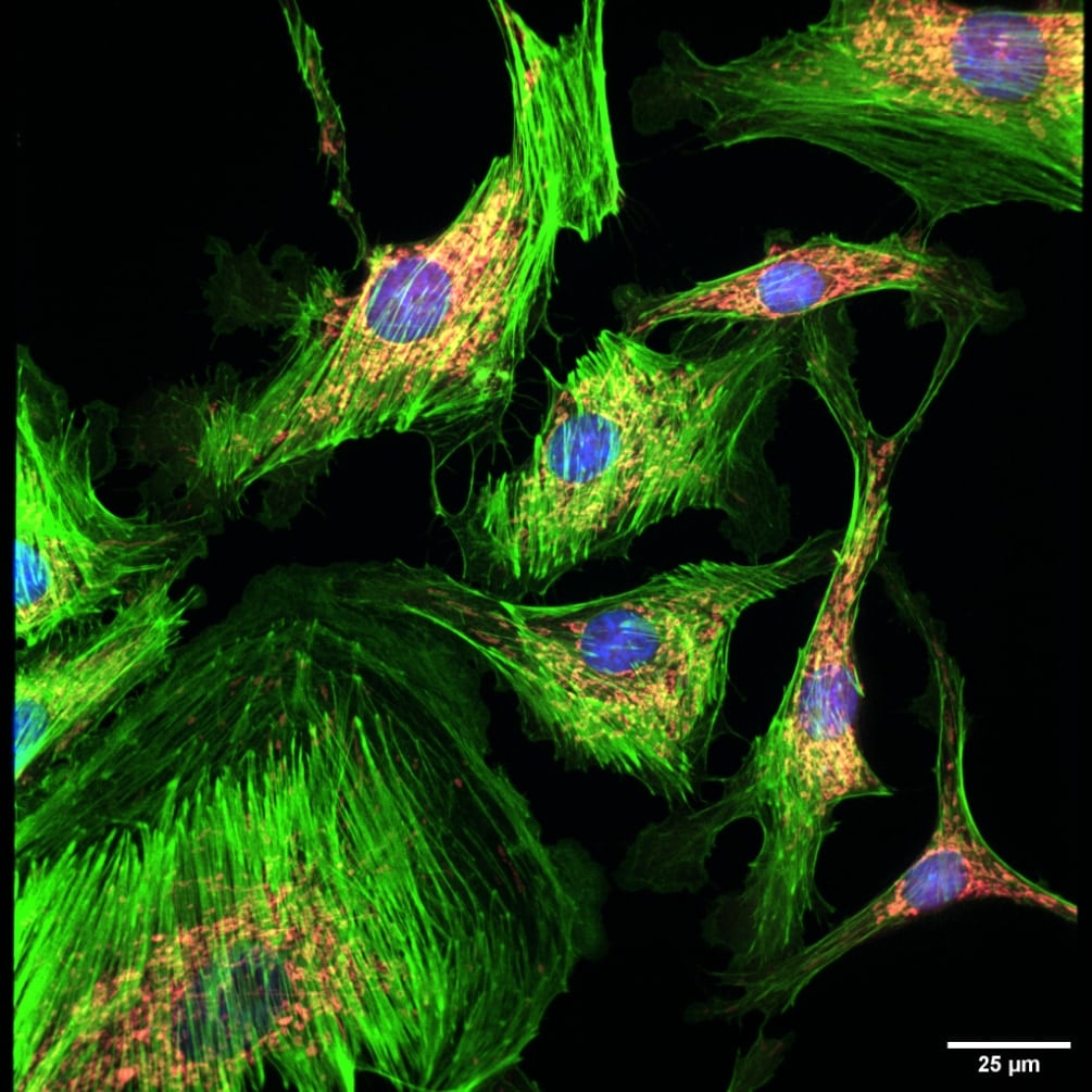
"After several years using the confocal system, we changed to RCM system with excellent results. The image acquisition is fast and with high resolution, which allow us to produce image stacks up to 200 slices without sample phototoxicity. We highly recommend RCM System for fixed and live-cell imaging"