RCM1
High contrast imaging. Low laser power.
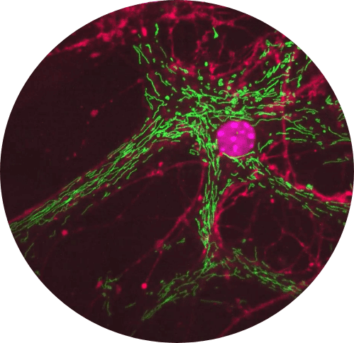
In this movie healthy mitochondria and fast-acting dynamics can be observed after long-term image acquisition
Super-resolution imaging.
Low laser power.
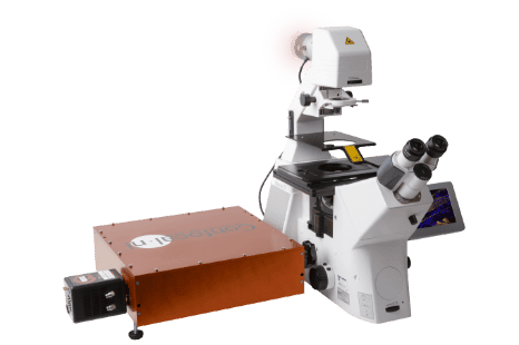
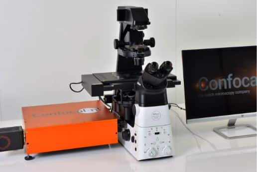
RCM1 is a highly sensitive super-resolution confocal system based on the Re-scan Confocal Microscope (RCM) principle. Capture datasets with 170nm raw resolution (120nm after deconvolution) by using 60x to 100x high numerical aperture (NA) magnification objectives and keep the laser intensity at a minimum. RCM1 acquires super-resolution images with only 85 nano-watts laser power.
How RCM1 improves your imaging experience
- Super-resolution confocal system
- High sensitivity
- Excellent add-on to any widefield microscope
- Easy to use
- API integration into third-party software
- VIS or NIR versions available
Discover the way RCM1 improves your imaging experience
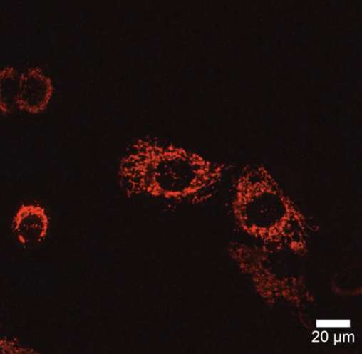
Long-term mitochondria imaging with no signs of phototoxicity.
Higher sensitivity and less phototoxicity with RCM1
RCM1 enables you to investigate live-cell dynamics in high resolution while keeping the laser power extremely low. Its high sensitivity makes RCM exceptionally suitable for challenging biological applications where the signal is limited.
RCM1-NIR version facilitates deep tissue biomedical imaging, achieving much higher sensitivity in the near-infrared wavelengths than other systems. It works with 640nm and 785nm wavelengths which, due to the longer wavelength, are less phototoxic and penetrate much better into biological samples.
Discover the way RCM1 improves your imaging experience
The benefits of using RCM1
Using low laser power increases cell survival and minimizes photobleaching during long-term time-lapses providing more relevant results. Unlike other super-resolution microscopy techniques, the RCM requires minimal light exposure for your samples!
Getting sharper and even higher contrast images, without averaging or integration, reduces the acquisition time and allows for a more precise analysis of the subcellular structures.
Discover the way RCM1 improves your imaging experience
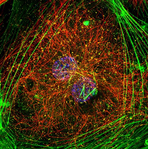
Hela cell stained for DNA (blue), Actin (green), Tubulin (red) and clathrin (yellow). Captured on RCM1 using a 100x 1.45 objective. Sample courtesy: Nicolas Touret, University of Alberta, Canada.
Applications for RCM1
Obtain super-resolution confocal images with standard dyes and without complicated training. RCM1 is an easy-to-use and versatile system, great to add to any microscopy facility. It’s a fruitful solution for individual research labs to easily convert any existing widefield fluorescence microscope into a super-resolution and highly sensitive confocal system. Record living cells and organoids in 4D super-resolution using very low laser power. Don’t worry about phototoxicity or photobleaching. RCM1 provides optimized conditions for live-cell imaging and facilitates posterior analysis with sharp and high contrast raw images.


RCM1: technical specs
RCM1 | |
|---|---|
Detector | Camera (sCMOS) |
Resolution in real time | 120 nm * |
Detector Sensitivity | up to 96% QE |
FOV | FN12: 150×150 μm (60x objective), FN18 when not using super resolution |
Optimized for super resolution | 100x, 60x (high NA) |
Speed | 1fps @ 512 x 512 pixels |
Wavelength | VIS or NIR |
Software | Micromanager, NIS Elements, Volocity |
Deconvolution with | Microvolution (real time), SVI Huygens |
Emission filters | standard quad band, optional external single band filter wheel |
* after deconvolution, raw image = 170nm

See for yourself how we improve your experience
Request your
personalized demo
- Your online demo will be shown in only 45-60 minutes
- Our experts will lead you through our products performance
- We will contact you and together, we pick a day and time

