Improved live cell imaging made accessible
Super-resolution. Low laser power. The solution for live cell imaging.
The fact that Confocal.nl allows for super-resolution in combination with very low laser intensity will be the winning strategy in live cell imaging.
Our solutions are as clear as our imaging
Sharper. Larger field of view. Easy to use.
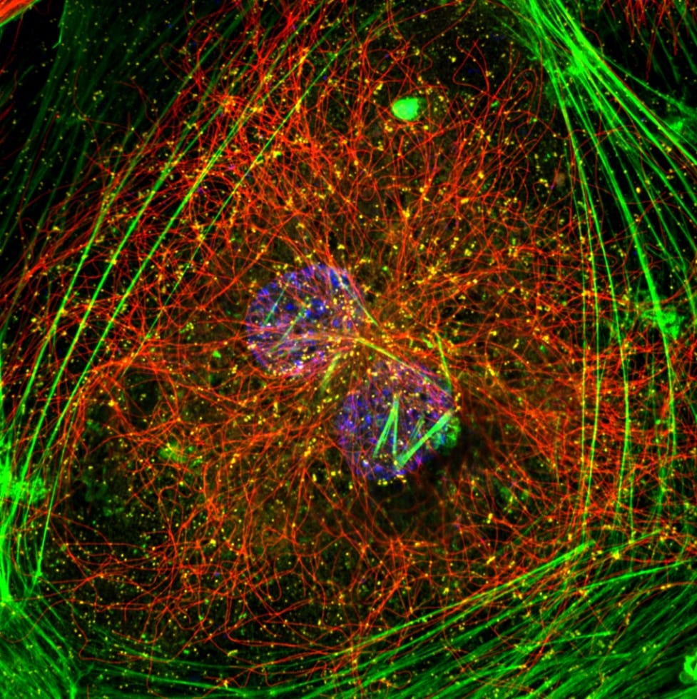
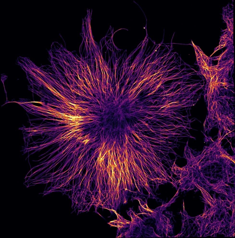
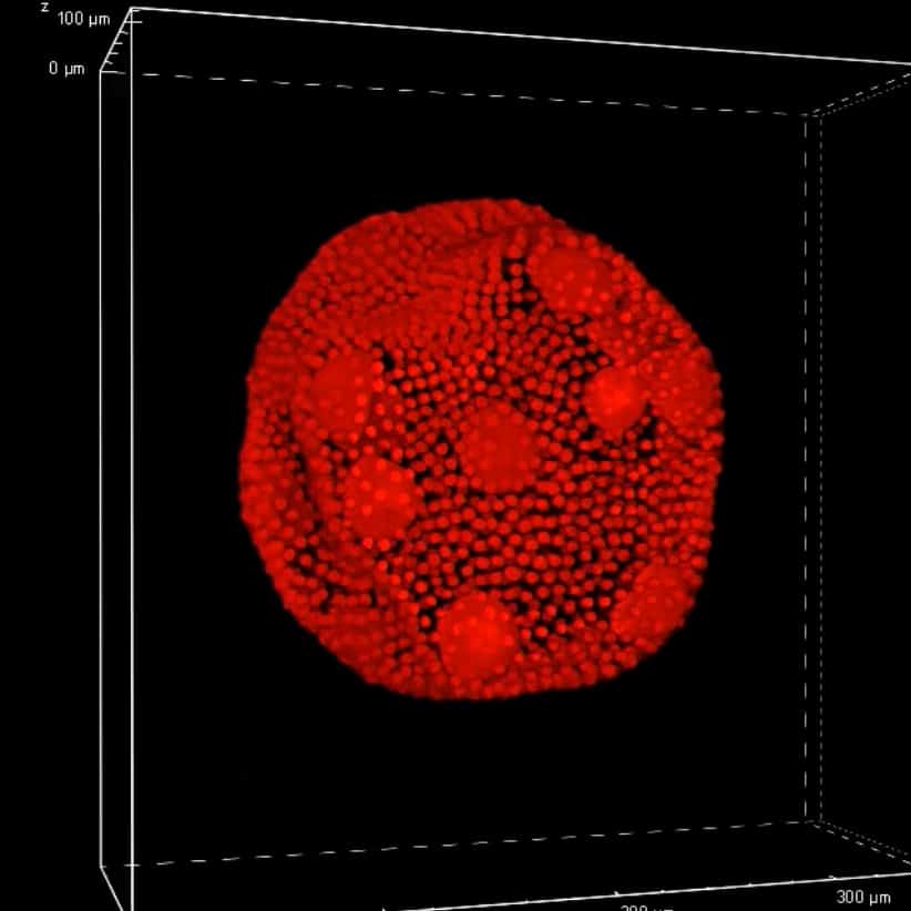
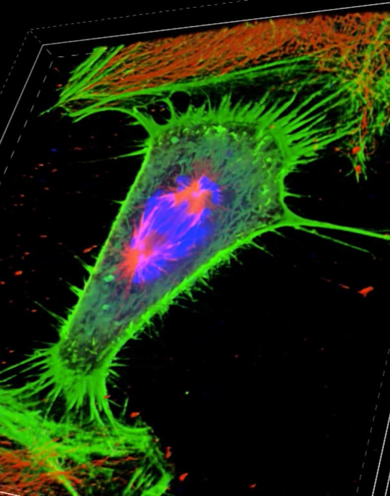
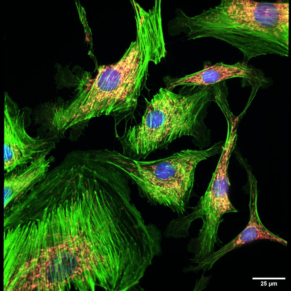
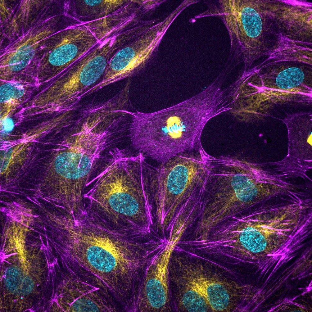
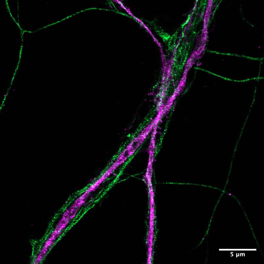
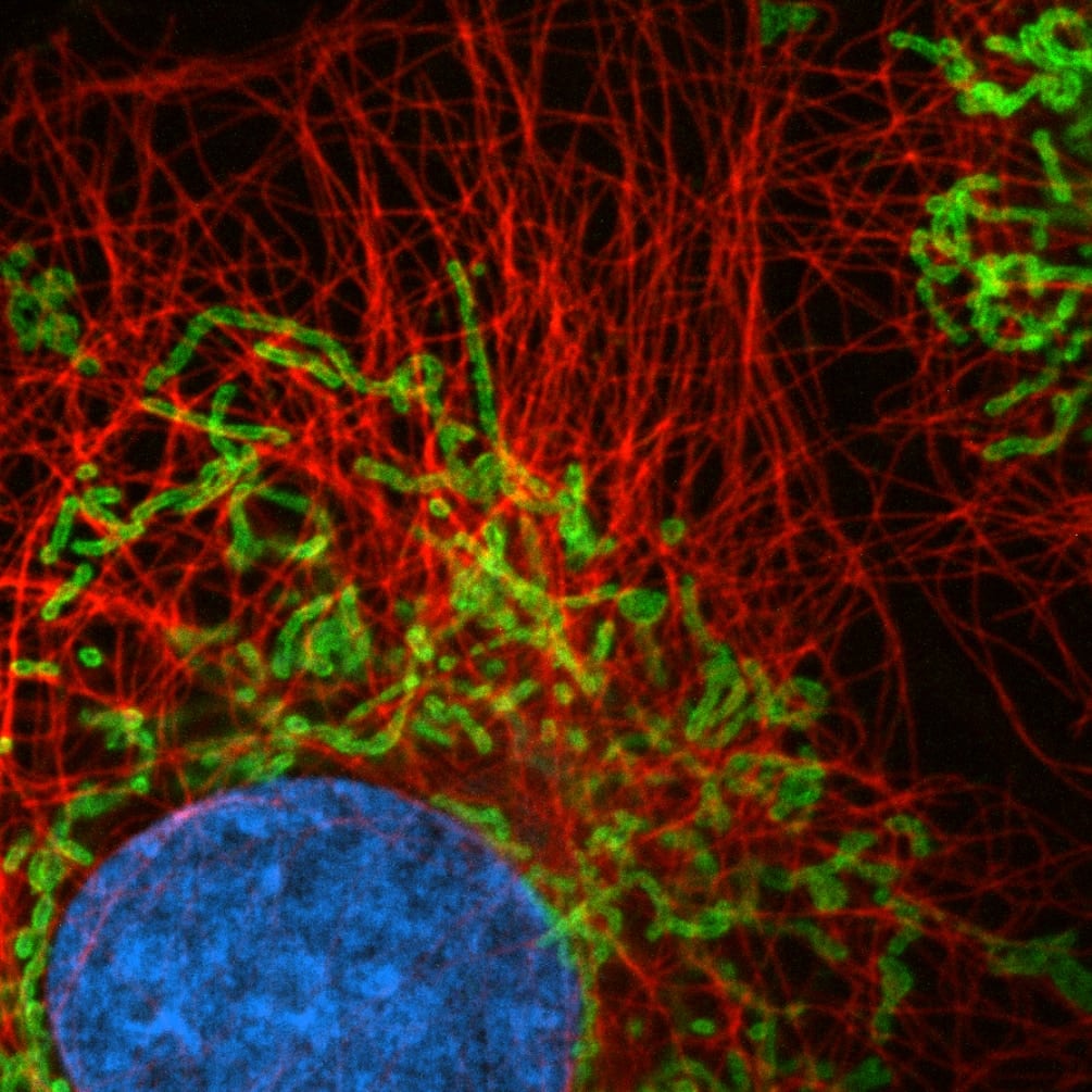
Sharp images, a large field of view and easy to use? It’s possible with Confocal.nl. Get more from your samples, with a higher resolution and a higher sensitivity than conventional confocal microscopes and study live samples longer. Our Re-scanning Confocal Microscope (RCM) uses multiple laser pointers and a camera as a detector. The low laser power required is live cell friendly: it prevents phototoxicity in your live samples and photobleaching of your fluorophores. Creating long-term time lapses at a super-resolution was never this accessible.
Want to discover Confocal.nl solutions for yourself?
Get an online demo now and discover how the products perform live in 45 to 60 minutes.
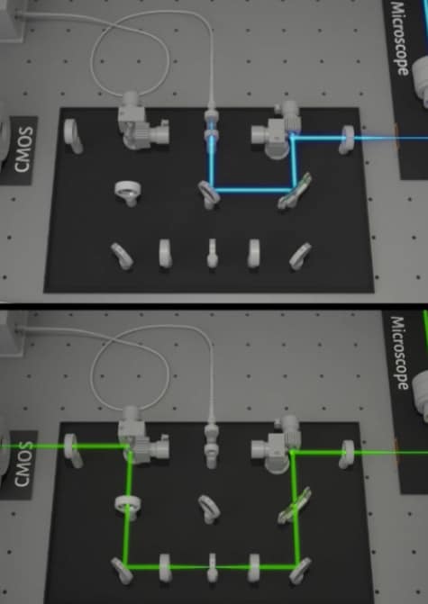
How we improve your imaging experience
Camera-Based Confocal Technology
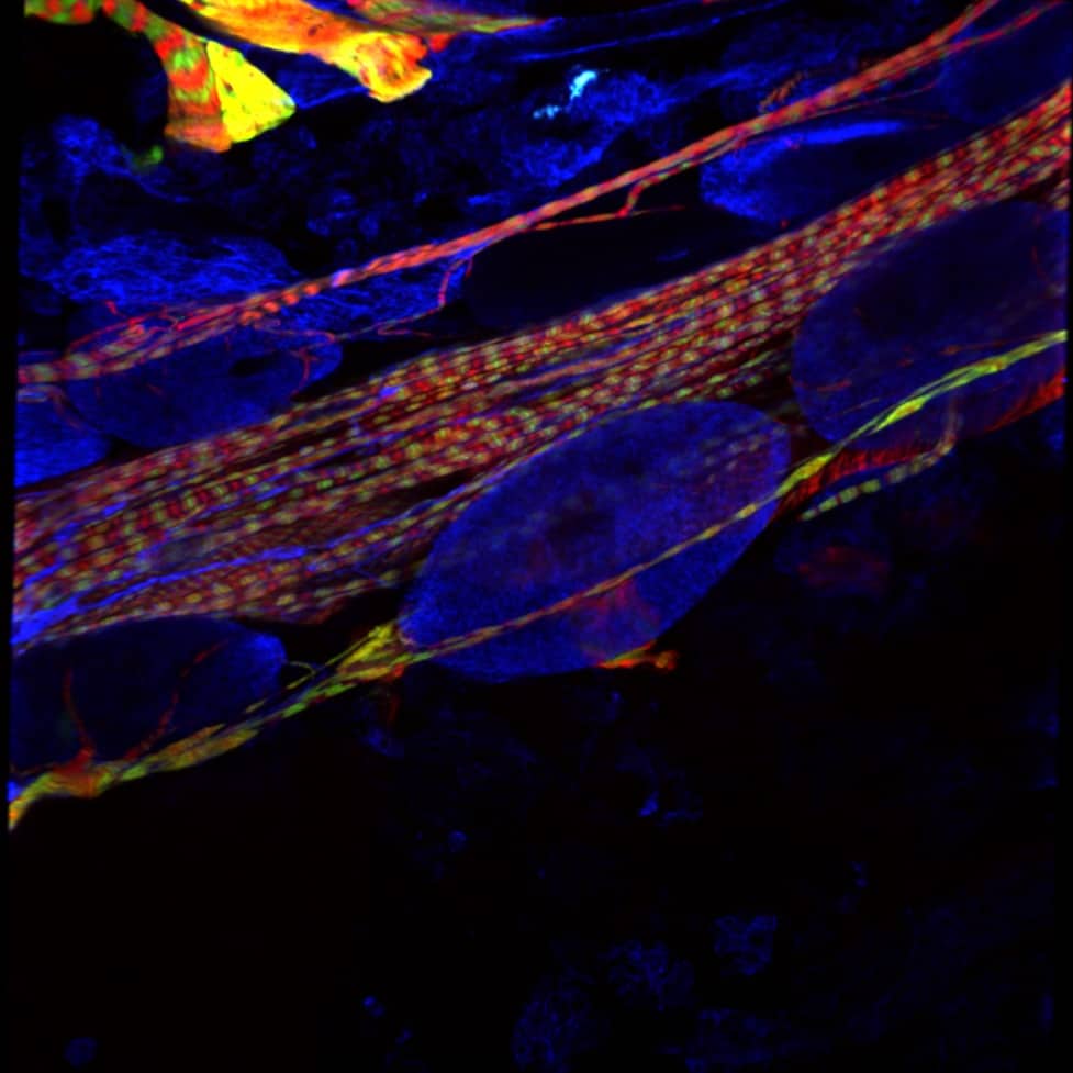
Re-scan Confocal Microscopy (RCM) is a new super-resolution technique, based on standard confocal microscopy. It’s extended with an optical re-scanning unit that projects the image directly on a CCD- or sCMOS camera. By using a sensitive camera as detector, the signal-to-noise ratio of the RCM is 4 times higher than in standard PMT-based confocal microscopes. This new technology improves lateral resolution and strongly increases sensitivity, while maintaining the sectioning capability of a standard confocal microscope. Closing down the pinhole is no longer necessary to increase resolution thanks to the re-scan step.
It is excellent for biological applications where a combination of super-resolution and high-sensitivity is required.
Learn all about improving you imaging
Confocal.nl in the news
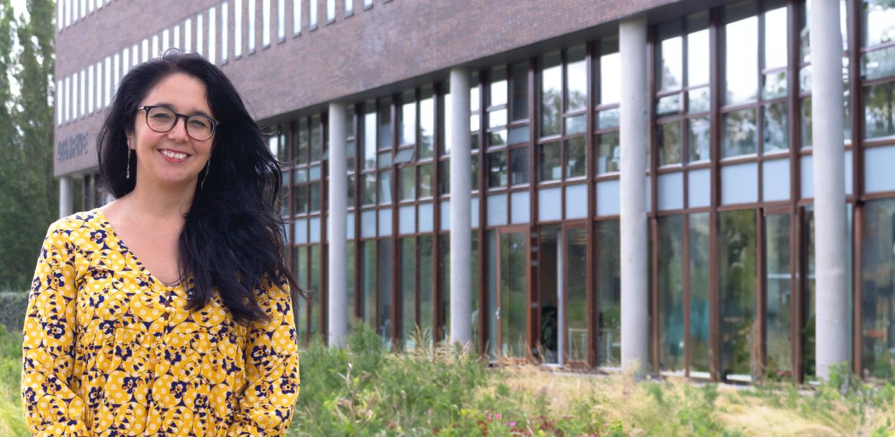
From Chile to the Netherlands: how Desiree started at Confocal.nl
Read more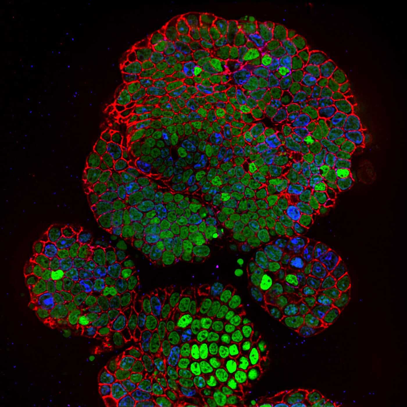
News
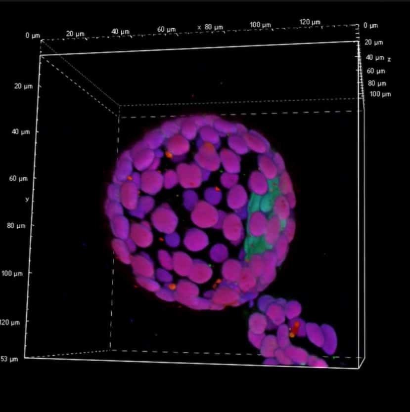
Line REscan confocal microscope NL5+: a fast and sensitive technique for studying organoids
Read more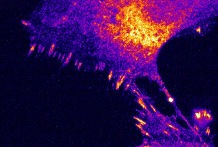
Articles
How super-resolved fluorescence microscopy led to a Nobel Prize
Read more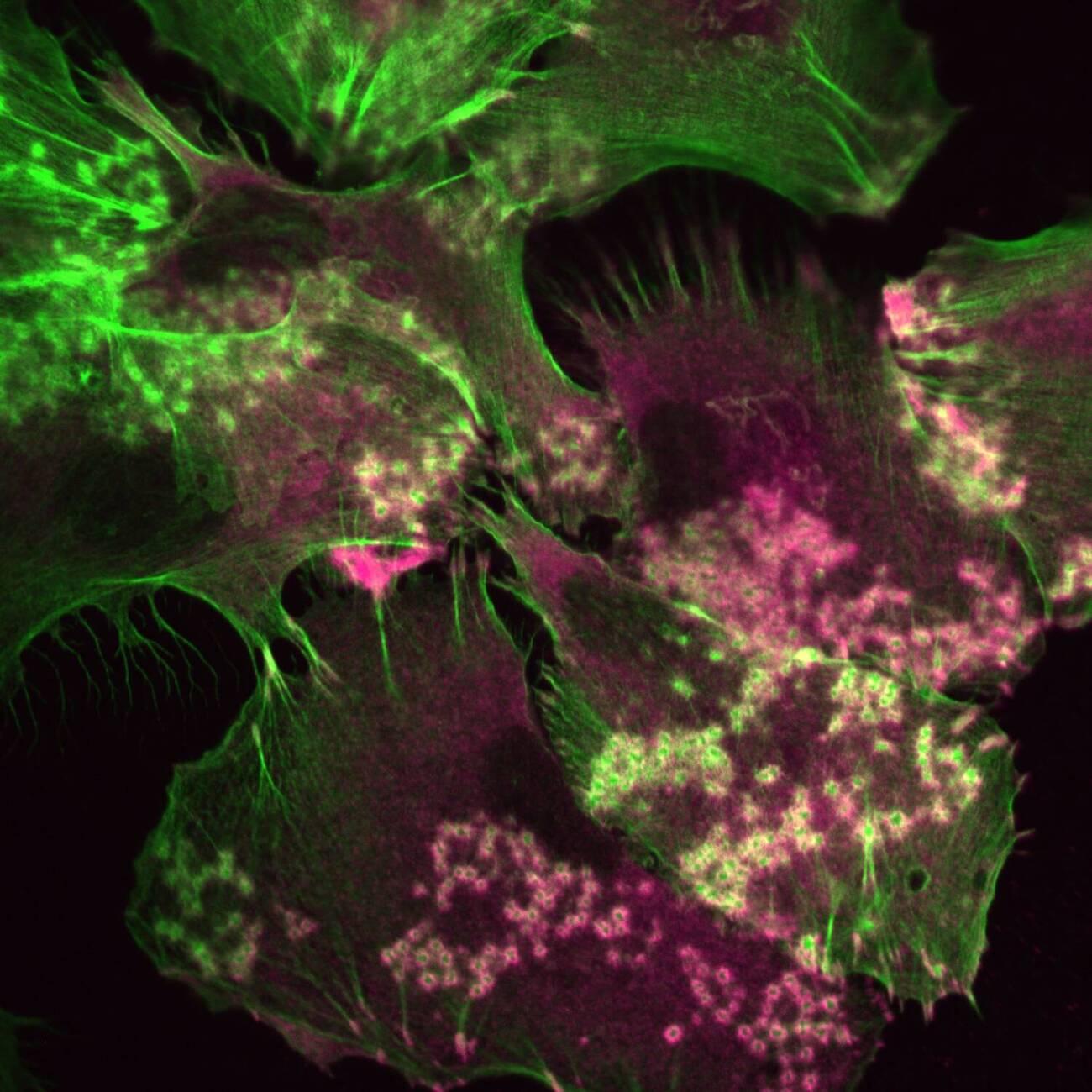
News
Confocal.nl enters new growth phase with investor and partner ECFG
Read moreDiscover our solutions
Ingenious design for the best live cell imaging you can get
RCM1
RCM2
RCM2.5
NL5
NL5+
NL5+
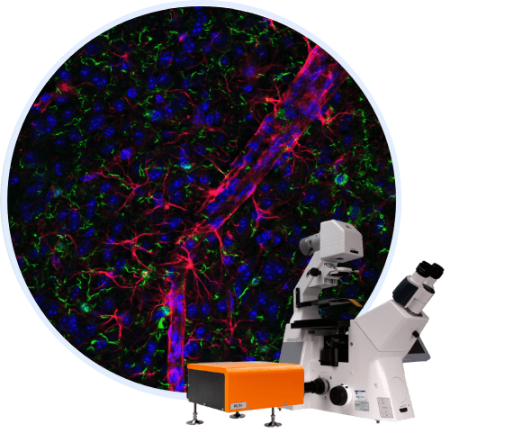
NL5+ has an additional filter, placed on top of your confocal system. Get high-contrast images from thicker specimens and create long time-lapse experiments for the imaging of low-signal samples.
RCM2
RCM1

RCM2.5
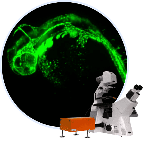
NL5
We kept cells alive for 61 hours under a microscope
The community challenged us
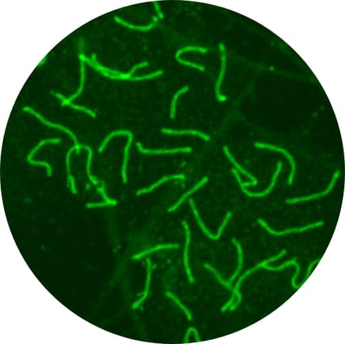
We took the challenge of long term live cell imaging and this is the result: 61 hours of live cell imaging without phototoxicity. HO1N1 cells expressing M itochondria-RFP through the Bacmam expression system. One image was taken every 10 seconds for 61 hours (giving a total of almost 22.000 images!). Laser power was measured to be 1 microwatt at the sample plane.
Movie taken by Confocal.nl, sample courtesy Dandan Ma (ACTA, Amsterdam), equipment provided by Marko Popovic (Nikon Center of Excellence, Amsterdam University Medical centers, VUmc, Amsterdam.)
Become a partner
Start working on the future of microscopy
As a partner of Confocal, you’ll be working on the future of microscopy alongside a young and dynamic team. We’ll work together to create the best imaging solutions and to distribute them. Let’s make RCM accessible across the world.
Distibrutors
Partners


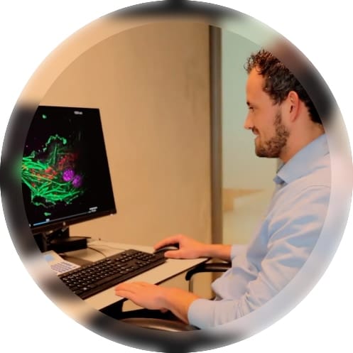
A team of enthusiastic microscopists
We improve your imaging experience
Founded in 2016, Confocal.nl strives to improve the microscopic imaging experience of all researchers. With our international and open-minded team, we develop innovative and ingenious designs in cooperation with our users, we provide the most user-friendly upgrade technology for microscopes. This brings opportunities to researchers to deliver breakthrough science. With the passion we have for science we create value for all our stakeholders.

See for yourself how we improve your experience
Request your
personalized demo
- Your online demo will be shown in only 45-60 minutes
- Our experts will lead you through our products performance
- We will contact you and together, we pick a day and time

